In April 1953, Francis Crick and James Watson published a paper in Nature which established the structure of DNA. Their paper was of the most significant discoveries in all of biology, giving rise to the field of molecular biology, while answering the question of how life reproduces itself.
The Hunt for the Hereditary Material

In the middle of the 19th century, the Austrian monk Gregor Mendel conducted a series of experiments with pea plants where he established some fundamental principles of hereditary. His pioneering work laid the groundwork for the field of genetics. However, the physical basis for the hereditary material of life remained a mystery for the time being.
In the early 20th century, the search for the hereditary material intensified. The American geneticists Thomas Hunt Morgan conducted important research at Columbia University with the fruit fly Drosophila that advanced our understanding of the role of chromosomes in heredity. Chromosomes were discovered in 1888, four years after the rediscovery of Mendel’s work on pea plants, and it was suspected that they were involved in the passing of hereditary traits. It was observed that patterns of trait transmission corresponded with the movement of chromosomes, an idea became known as the Chromosomal Theory of Inheritance. But in the early 20th century it wasn’t clear how to prove this.
Morgan and his team conducted a breeding and crossbreeding program involving millions of fruit flies, and through this program was able to work out correlations between particular traits and individual chromosomes that showed chromosomes were involved in the passing of genetic material. More specifically, Morgan was able to establish that one of the mutations he found in his fruit flies, a change in eye color, only occurred in males. This could only be explained if the mutation occurred on the male Y chromosome, which Morgan was also able to establish.
Chromosomes are made up mostly of proteins, with a bit of nucleic acid. The search was on the discover the molecule on the that transmitted hereditary material, and the early assumption was that it was a protein. By the 1940s various biological techniques were devised to break down the different biological materials – proteins, lipids, carbohydrates, and nucleic acids – and only when nucleic acids were broken down was the hereditary material not transmitted. However, there was a bias towards assuming that proteins were the hereditary material and more evidence was needed to convince the scientific community.
This evidence came from the experiments of Alfred Hershey and Martha Chase in the early 1950s. They designed an elegant set of experiments using radioactive tracers in viruses. This was accomplished by growing viruses in a solution with different radioactive isotopes, one with radioactive sulfur (a main component of proteins but not found in DNA) to label the protein and one with radioactive phosphorous (a main component of DNA but not found in proteins) to label the DNA. They allowed the labeled viruses to infect a culture of E. coli bacteria and initiate reproduction and then were able to trace which material was responsible for hereditary transmission by a process called centrifugation. The results of this experiment produced strong and convincing evidence that DNA, and not proteins, carried the hereditary material.
This laid the groundwork for the quest of understanding deoxyribonucleic acid or DNA which was now shown as the physical material responsible for carrying hereditary information.
Unraveling the Structure of DNA
By this point in the middle of the 20th century, it was realized by most biologists that a detailed knowledge of the three-dimensional structure of DNA was critical to understanding how the molecule works. This is the classic biological principle that form follows function. Many people were racing towards this inevitable discovery, and indeed Crick and Watson’s work was dependent on the work of others, most notably Rosalind Franklin and her X-ray crystallography. Franklin took a purified DNA crystal and produced the clearest X-ray crystal image to date. On a visit to London, James Watson was able to view this image and working with Crick they were able to deduce some important structural elements from it. First, they learned that the molecule had to form some type of helix. Second, they were able to figure out the width of this helix, which was about two nanometers, or about the size of one nucleotide.
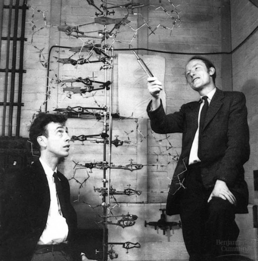
The was one other piece of information was important to determining the structure of DNA. It was observed through experiments that there was a consistent ratio between the nitrogenous bases of the DNA. The amount of adenine always equaled the amount of cytosine, and the amount of guanine always equaled the amount of thymine.
Working with this information, Watson and Crick began building literal models of DNA strands. Through a series of trial and error, they eventually were able to build a model that fit with all of the data. Their model is the famous double helix.
The double helix model that Crick and Watson established contained a few major features. The outside consists of two sugar-phosphate backbones held together by hydrogen bonds, with nitrogenous bases on the inside. There are four types of bases – adenine (A), cytosine (C), guanine (G), thymine (T), with pairing always occurring with A & T and C & G. The copying mechanism works with the helix “unzipping” into two separate strands that act as templates for a new molecule due to the pairing mechanism.
Continue reading more about the exciting history of science!